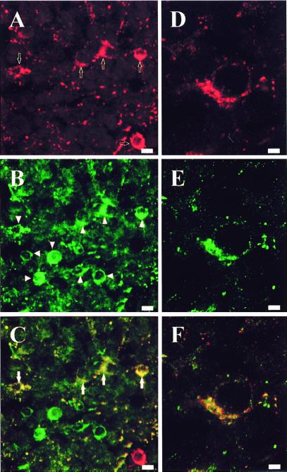
Figure 2.
Dual-channel confocal laser scanning microscope photomicrographs comparing the distribution of 3β-HSD and GABAA receptor β2/β3 subunit-like immunoreactivity in the frog hypothalamus. (A–C) Frontal section through the posterior tuberculum labeled with the antiserum against 3β-HSD revealed with DARS/TXR (A; arrows) or the monoclonal antibody against the β2/β3 subunits revealed with GAMS/Alexa-488 (B; arrowheads). Combination of the two images acquired in A and B revealed the presence of β2/β3 subunit-like immunoreactivity in a subset of 3β-HSD-positive neurons (C; arrows). (Bars = 10 μm.) (D–F) Frontal section through the nucleus of the periventricular organ labeled with the antiserum against 3β-HSD revealed with DARS/TXR (D) or the monoclonal antibody against the β2/β3 subunits revealed with GAMS/Alexa-488 (E). Combination of the two images acquired in D and E revealed the coexistence of 3β-HSD- and β2/β3 subunit-like immunoreactivity in the same neuron (F). (Bars = 2 μm.)

