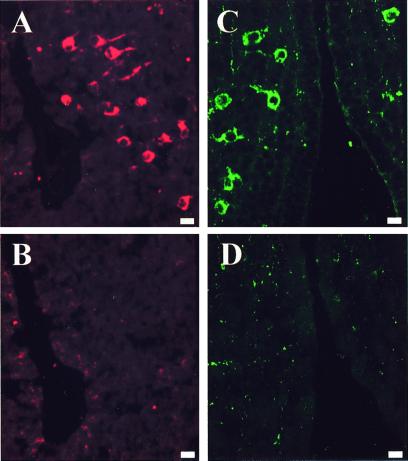
Figure 3.
Labeling of consecutive coronal sections through the anterior preoptic area, showing the specificity of the immunocytochemical reactions. (A and B) Specificity control of the 3β-HSD immunostaining. Adjacent sections were incubated with the antibodies against 3β-HSD (A) or with the antibodies preabsorbed with purified human type I 3β-HSD (10−6 M) (B). (C and D) Specificity control of the α3 subunit immunostaining. Adjacent sections were incubated with the antibodies against the α3 subunit (C) or with the antibodies preabsorbed with the purified peptide hapten (10−6 M) (D). (Bars = 10 μm.)

