Figure 1.
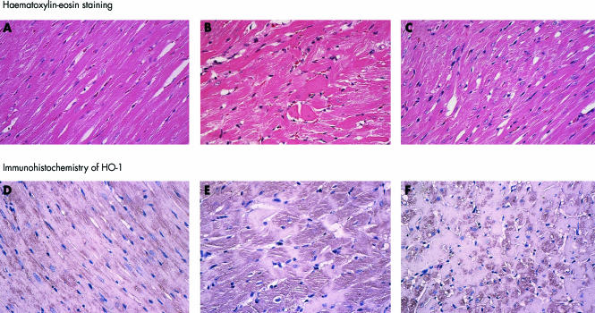
Light micrographs. Upper panels (A, B, C): haematoxylin–eosin staining. Lower panels (D, E, F): immunohistochemistry for HO-1 (polyclonal antibody against haem oxygenase-1). The LETO rats showed normal morphology (A). In the LETO rats, immunoreactivity against HO-1 antibody was slight (D). In the OLETF rats, hypertrophied cardiomyocytes, disarray of myofibres, and scarcity of myofibrils were observed (B). HO-1 was increased in the cytoplasm of cardiocytes (E). All the above changes were smaller in the OLEFT+ARB subgroup (C and F). Magnification ×100. ARB, angiotensin II receptor blockade.

