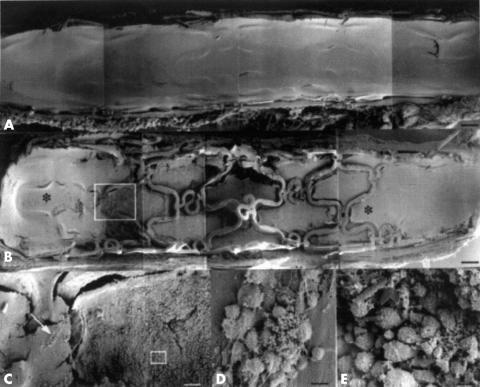
Figure 2.
Low power scanning electron micrograph of control stent (A) and 48 μCi stent (B) at 12 months. The entire control stent is endothelialised, whereas the 48 μCi stent is endothelialised at the ends (* in B). Parts of the central portion of the 48 μCi stent are non-endothelialised, and bare or partially covered struts are present. The transition zone between endothelialisation and non-endothelialisation is outlined in B and shown at medium power in C. Two areas are further magnified (arrow and box in C corresponding to D and E, respectively) to show adherent platelets and inflammatory cells on partially endothelialised (D) and non-endothelialised (E) parts of the stent. Bars = 0.5 mm in A and B, 0.1 mm in C, 0.01 mm in D and E. Reproduced from: Farb A, et al. Circulation 2001;103:1912–9, with permission of the American Heart Association.

