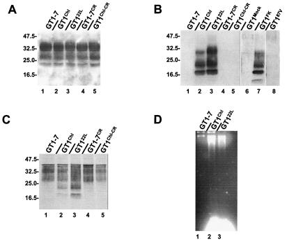
Figure 1.
Cell lysates of GT1 lines were loaded onto 12% polyacrylamide gel, transferred onto poly(vinylidene difluoride) membrane, and PrPC detected with antibody P45–66 raised against the amino terminus of the protein (A) or with a mixture of mAb (SAF 60, SAF 69, and SAF 70) raised against the carboxy terminus of the protein (C). PrPSc was detected with the same mix of mAbs but after proteinase K digestion (B). Molecular mass markers are indicated on the left in kDa. In D, DNA fragmentation was demonstrated in infected cells by the DNA laddering technique.

