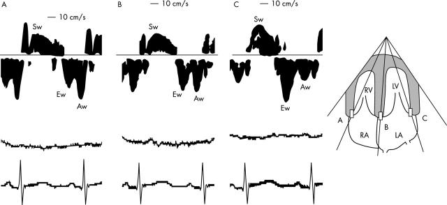
Figure 1.
Representative recordings of the velocity spectrum of the wall motion assessed by pulsed wave Doppler tissue echocardiography in a baby two days after birth in the apical four chamber view. Recordings from (A) the tricuspid annulus, (B) the base of the ventricular septum, and (C) the mitral annulus. The corresponding ECG and respiration curve were simultaneously recorded. Aw, peak atrial systolic motion velocity; Ew, peak early diastolic motion velocity, LA, left atrium; LV, left ventricle; RA, right atrium; RV, right ventricle; Sw, peak systolic motion velocity.

