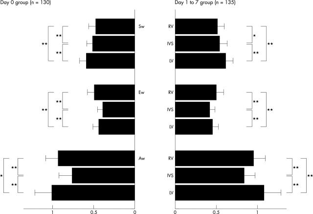
Figure 2.
Ratio of pulsed wave Doppler tissue velocities at three ventricular walls of neonate groups to those of the children group. The PWDT velocity at three ventricular walls in each phase of the cardiac cycle of each neonate was expressed as a ratio to the mean value of peak PWDT velocity of the children group. RV was recorded from lateral site of the tricuspid annulus, IVS (interventricular septum) from its base, and LV from the lateral site of the mitral annulus. *p < 0.0005, **p < 0.0001.

