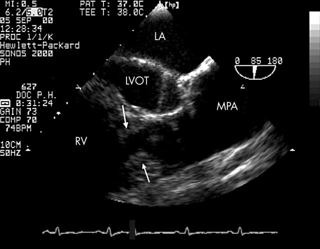
Figure 1.
Double chambered right ventricle (DCRV). High and discrete obstruction of the right ventricular cavity (arrows) adjacent to the pulmonary valve, demonstrated by transoesophageal echocardiography in the longitudinal plane (85°). In this case DCRV is associated with valvar pulmonary stenosis. LA, left atrium; LVOT, left ventricular outflow tract; MPA, main pulmonary artery; RV, right ventricle.

