
Figure 3.
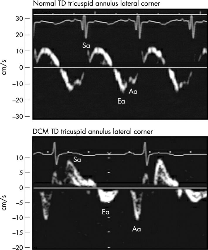
TD at the lateral tricuspid annulus in a patient who subsequently required transplantation (lower panel) compared with a normal control participant (upper panel). Note the significantly diminished tricuspid Ea velocity measuring 5 cm/s in the patient requiring transplantation. There is partial fusion of the Ea (16 cm/s) and Aa velocity in the normal control participant.