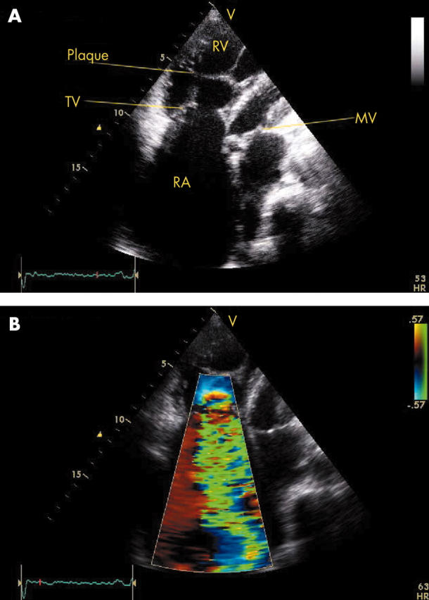
Figure 2.

(A) Transthoracic echocardiogram in a modified apical four chamber view showing thickened and rigid tricuspid valve (TV) leaflets in an abnormal, fixed open position during systole. Endocardial plaque formation of the subvalvar apparatus is also notable. (B) Colour flow Doppler imaging in the same view shows severe tricuspid regurgitation through a wide regurgitant orifice. MV, mitral valve; RA, right atrium; RV, right ventricle.
