Figure 5.
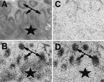
sst3 in human lymphoreticular tissue. (A) Hematoxylin-eosin-stained section showing parts of a human tonsil containing a T-cell-rich interfollicular area (around the asterisk) and B-cell-rich germinal centers (arrows). (B) Autoradiogram showing total binding of 125I-[LTT]-somatostatin-28, with labeling of the interfollicular area (around the asterisk) and of germinal centers (arrows). (C) Autoradiogram showing nonspecific binding of 125I-[LTT]-somatostatin-28 (in the presence of 10−6 M somatostatin-28). (D) Autoradiogram showing binding of 125I-[LTT]-somatostatin-28 in the presence of 10−7 M compound 10. Binding to the interfollicular area is abolished, strongly indicating sst3 expression in this area. Binding to the germinal centers remains; they are known to express sst2 (43).

