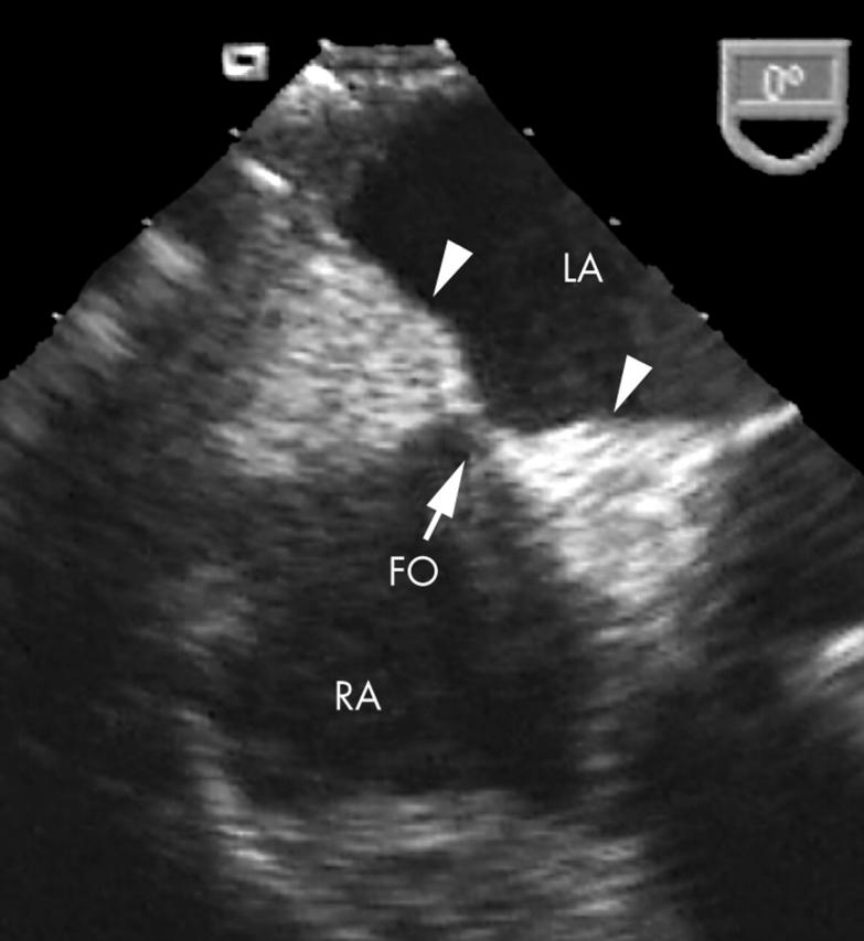
Figure 2.

Transoesophageal echocardiogram at the mid-oesophageal level showing the classic hour glass shaped lipomatous hypertrophy of the intra-atrial septum (arrow heads). Note the clearly defined septal borders along with the bright echogenicity of thickened tissue, which clearly spares the fossa ovalis (FO, arrow).
