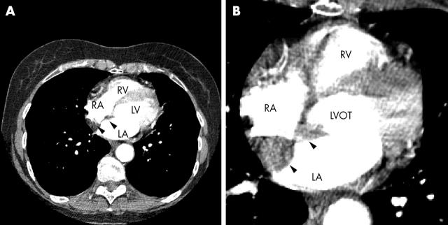
Figure 4.
Contrast enhanced multislice computed tomography (CT) of the thorax showing thickening of the intra-atrial septum (arrow heads). The axial slices of the body show the heart in an approximate four chamber view. (A) Lower cardiac section; (B) close up of a higher cardiac section. The contrast agent generated a high attenuation in the LV, LV outflow tract (LVOT), LA, RV, and RA. The septal thickening is clearly visualised. Note also that CT allows characterisation of the nature of the septal tissue, which is clearly hypointense to normal myocardium and has an attenuation similar to subcutaneous fat.

