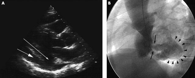
Figure 1.
(A) Echocardiogram in the parasternal long axis view showing a defect in the posterior-inferior left ventricular wall (small arrow) and posterior pericardial effusion (large arrow). (B) Ventriculogram in the left anterior oblique view showing a bulge in the posterolateral wall of the left ventricle (arrowheads) connected to the ventricular chamber by a narrow well defined neck (arrows).

