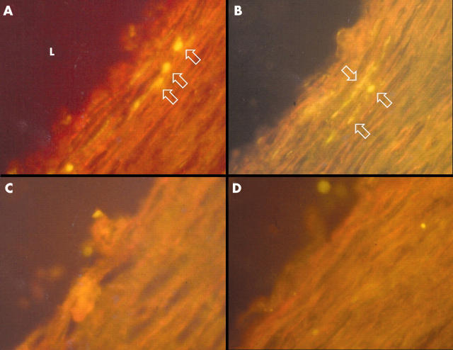
Figure 1.
Immunohistochemical staining of 5 μm sections of a coronary endarterectomy specimen with monoclonal antibodies to two distinct antigens of Chlamydia pneumoniae. Sequential sections stained with (A) fluorescein isothiocyanate (FITC) labelled C pneumoniae major outer membrane protein antibody (clone RR402) and (C) an IgG3 isotype control antibody. Sequential sections of the same specimen stained with (B) FITC labelled C pneumoniae heat shock protein-60 antibody (clone A57-B9) and (D) an IgG1 isotype control antibody. Arrows indicate positively stained cells. L, artery lumen. (Original magnification ×400).

