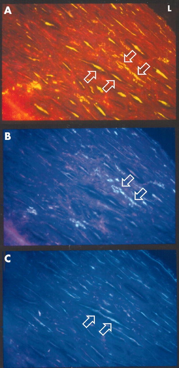
Figure 3.

Double immunohistochemical staining of 5 μm sections of a coronary endarterectomy specimen showing smooth muscle cells and macrophages expressing MMP-9. (A) FITC staining of MMP-9. (B) AMCA staining of CD68. (C) AMCA staining of α smooth muscle cell actin. Upward arrows indicate cells positive for α smooth muscle cell actin and MMP-9 stain. Downward arrows indicate cells positive for CD68 and MMP-9 stain. (Original magnification ×400).
