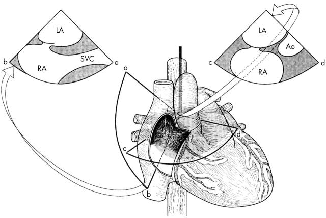
Figure 3.
Schematic drawing demonstrating the anatomy of the interatrial septum and the potential superiority of using a more vertical plane (sector image to the left). See text for details. Ao, aorta; LA, left atrium; RA, right atrium; SVC, superior vena cava. Adapted from Chenzbraun et al. J Am Soc Echocardiogr 1993;6:417.

