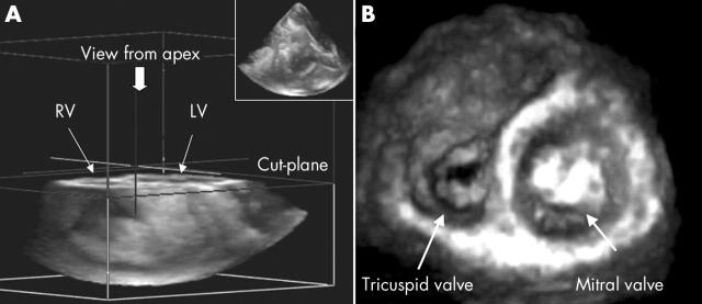
Figure 1.
Using four ECG triggered cardiac beats with the patient breath holding, four subvolumes were time aligned to render a pyramidal shaped full volume image (panel A, top right corner). To obtain the tricuspid valve area from the ventricular perspective, the three dimensional full volume was cropped using the elevational plane from the apex.

