
Figure 12.
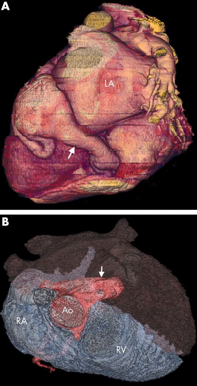
(A) Three dimensional VR (posterior view) of the circumflex artery fistula showing major dilatation of the receiving vessel (the coronary sinus; arrow). (B) This VR superior view helps to show the LCA fistula (arrow) in the RA and subsequent dilatation of the right heart chambers.