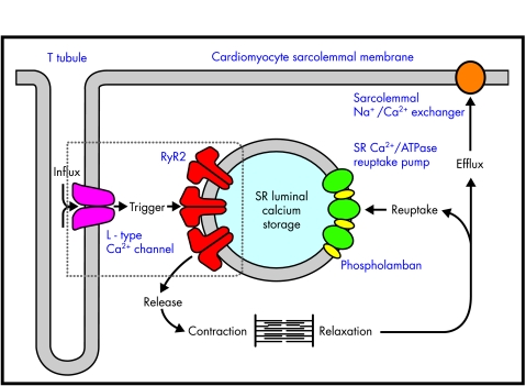
Figure 1.
Cardiomyocyte excitation–contraction coupling. Major components of E-C coupling within the myocyte are shown. The movement of Ca2+ around the cell is indicated by bold arrows. Note the close association of sarcoplasmic reticulum RyR2 with the sarcolemmal L type voltage dependent channel (box), allowing localised calcium induces calcium release events to take place. SR, sarcoplasmic reticulum.

