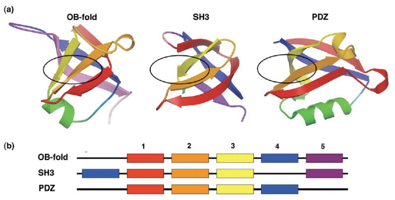
Figure 1.

Topologies of the OB-fold, SH3 domain, and PDZ domain superfolds. (a) The OB-fold is shown with the five β-strands of the central barrel colour-coded: strand 1 is red, 2 is orange, 3 is yellow, 4 is blue, 5 is lavender. The SH3 and PDZ domains have corresponding β-strands coloured as in (a). The approximate canonical ligand-binding sites each fold are indicated by black ovals, (b) A schematic illustrating the relationships of the β-strand secondary structure among the three superfolds, coloured as in (a). SH3 doman β-strand 4 is permuted to the N terminus relative to the OB-fold, while the PDZ domain lacks β-strand 5.
