
Fig. 1.
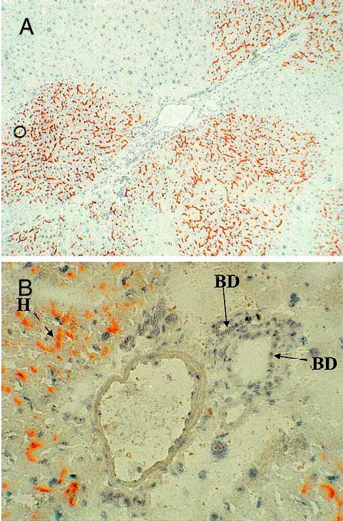
(A) Histochemical stain for DPPIV of the hybrid livers at 3 months after infusion of DPPIV-positive hepatocytes into DPPIV-negative rat liver, after treatment with retrorsine and partial hepatectomy. Positive stain result is indicated by the canalicular pattern of red/orange color. Variable portions of lobules are occupied by the DPPIV-positive cells. (B) Higher-power photomicrograph of a portal triad from a hybrid liver. Many hepatocytes (H) are positive for DPPIV, whereas biliary epithelium (BD) is uniformly negative.