
Fig. 2.
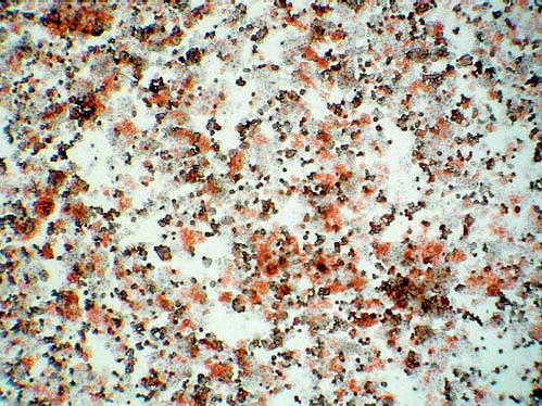
DPPIV stain of air-dried smear of freshly isolated hepatocytes from a hybrid liver. Positive cells are stained red/orange. Normally the stain is limited to the bile canaliculus, but after collagenase perfusion, the canalicular structures are disrupted and canalicular markers are seen over the entire hepatocyte membrane.19