
Fig. 5.
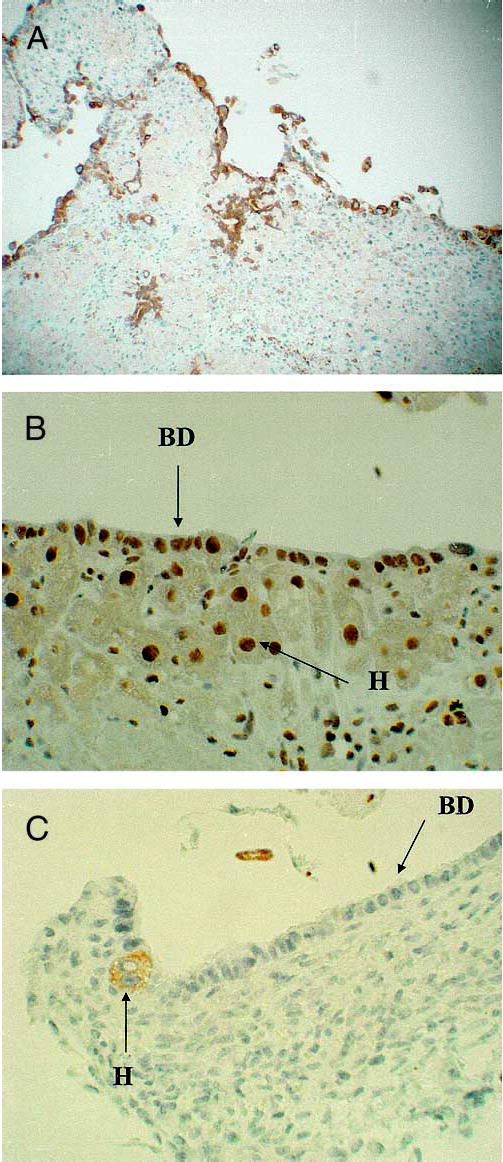
Organoid cultures from hybrid livers at day 21. (A) Immuno-histochemical stain for cytokeratin 19, a marker for biliary epithelium. Most of the surface biliary epithelium is positive for cytokeratin 19. (B) Immunohistochemical stain for PCNA. Most of the surface biliary epithelium and the underlying hepatocytes have positive (brown color) nuclei, indicating the cells are into the cell cycle. (C) Immunohistochemical stain for the hepatocyte marker HEPPAR shows that the biliary epithelium (BD) is negative for this marker. A single hepatocyte (H) adjacent to the surface biliary epithelium is positive for HEPPAR.