
Figure 2.
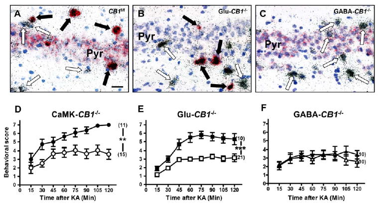
Deletion of CB1 in Glutamatergic Cortical Neurons, but Not in GABAergic Neurons, Increases Susceptibility to KA-Induced Seizures
(A–C) Micrographs showing double in situ hybridization of CB1 mRNA (red staining) together with GAD65 mRNA (silver grains) in the CA3 region of CB1f/f (A), Glu-CB1−/− (B), and GABA-CB1−/− mice (C). In (A), CB1 is present in both GABAergic interneurons and pyramidal neurons. In (B), CB1 is present only in GABAergic interneurons. In (C), CB1 is present only in pyramidal neurons. Blue staining, toluidine blue nuclear counter-staining. Filled arrows, GABAergic interneurons (GAD65-positive) expressing CB1 mRNA. Open arrows, GABAergic interneurons lacking CB1 expression. Bar, 25 μm. For similar analysis of CB1 expression in CaMK-CB1−/− mice, see Figure 2 of Marsicano et al. (2003).
(D–F) Seizure scoring (30 mg/kg KA) of conditional CB1 mutant mice (filled symbols), as compared to control CB1f/f littermates (open symbols). Seizures are worsened in CaMK-CB1−/− (D) and in Glu-CB1−/− (E), whereas they do not differ from wild-type controls in GABA-CB1−/− mice (F). In brackets, number of animals for each experimental group. Data are presented as mean ± SEM. **p < 0.005; ***p < 0.001 (by two-way ANOVA, factor genotype).