
Figure 4.
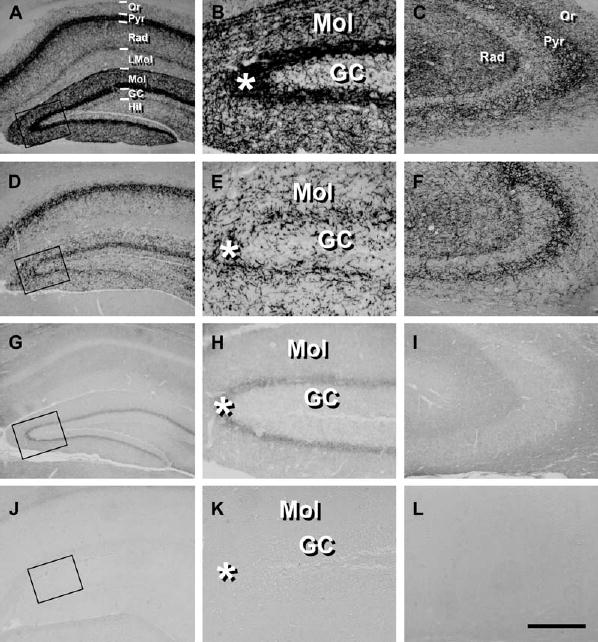
CB1 Protein Is Present in Glutamatergic Hippocampal Neurons
Micrographs showing the immunohistochemical analysis of CB1 expression in wild-type (A–C), CaMK-CB1−/− (D–F), GABA-CB1−/− (G–I) and complete CB1 knock-out mice (J–L).
(B, E, H, and K) Higher magnification micrographs of the areas enclosed in the square in (A), (D), (G), and (J), respectively.
(C, F, I, and L) Detail of the CA3 hippocampal region.
GC, granule cell layer of dentate gyrus; Hil, hilar region of dentate gyrus; LMol, stratum lacunosum-molecularis; Mol, stratum molecularis; Or, stratum oriens; Pyr, CA1/CA3 pyramidal cell layer of hippocampus; Rad, stratum radiatum. Asterisks indicate the inner third of the molecular layer. Bar, 100 μm (A, C, D, F, G, I, J, and L); 25 μm (B, E, H, and K).