
Fig. 5.
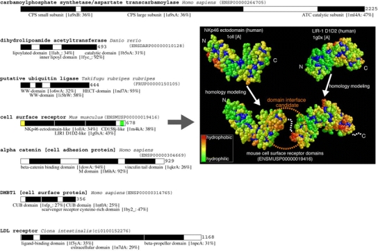
Eukaryotic ORFs with multiple model structures covering more than 70% of entire protein. In each of the bar representation of proteins, a black box is a region with 3D structure. A name and PDB ID of a template structure and amino acid sequence identity between template and target domains are given below the black box. A yellow box is a putative signal peptide and green box is a putative transmembrane region. Template and modeled structures of ENSMUSP00000019416 were shown on the right side of the figure. Each domain is colored by hydrophobicity. A hydrophilic residue is in green and a hydrophobic residue is in red. A buried residue is in deep blue