
Figure 2.
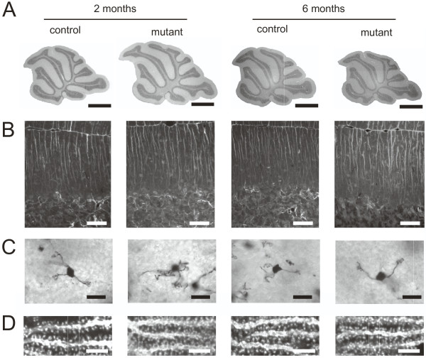
Cerebella from mutant and control animals look indistinguishable at the age of two and six months. (A) Normal lobulation pattern. Sagittal paraffin sections stained with hematoxylin/eosin, scale bar = 1 mm. (B) Bergmann glia are normal and no astrogliosis is evident. GFAP stained paraffin sections (lobule 8); scale bar = 50 μm. (C) Granule cell dendrites are normally developed. Golgi impregnated sections showing individual granule cells; scale bar = 20 μm. (D) Purkinje cell spines are normal; single confocal planes of Purkinje cell dendrites stained for calbindin, scale bar = 5 μm.