
Figure 3.
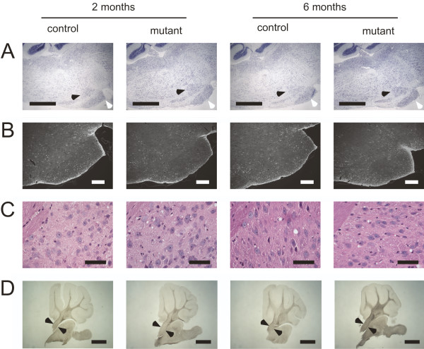
The pontine nuclei from mutant and control animals look indistinguishable at the age of two and six months. (A) The pontine nuclei (white arrow head) and the reticulotegmental nucleus of the pons (black arrow head) appear normal (Nissl stained paraffin sections), scale bar = 1 mm. (B) No astrogliosis is evident on paraffin sections stained for GFAP, scale bar = 200 μm. (C) The neurons in the pontine nuclei appear normal by hematoxylin eosin stain, scale bar = 50 μm. (D) The middle cerebellar peduncle (between arrow heads), the tract carrying the axons from the pontine nuclei to the cerebellar hemispheres, appear normal in size (unstained 50 μm sagittal sections), scale bar = 1 mm.