
Figure 4.
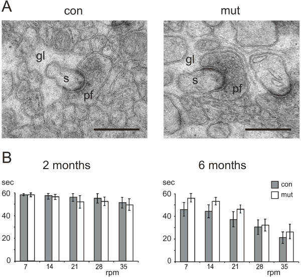
Synapses appear normal. (A) The ultrastructure of parallel fiber to Purkinje cell synapses was normal by electron microscopy between 2 months old control (con) and mutant (mut) animals. Parallel fibers (pf) synapse on Purkinje cell spines (s) that are fully enwrapped in Bergmann glial processes (gl). Scale bar = 500 nm. (B) Mutants performed normally on a Rotarod. The test was performed as described [27] at fixed speed levels (rpm: revolutions per minute) at the age of two and six months. The latency to fall was measured in seconds (sec) and the experiment was terminated after 60 s. The mean +/- (standard error) is shown (n>10 animals per group).