
Figure 4.
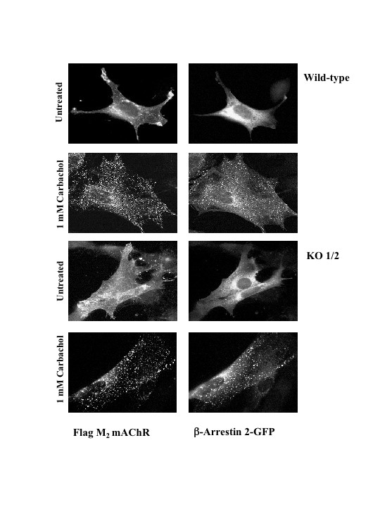
Stimulation of M2 mAChRs leads to stable co-localization of β-arrestin 2-GFP at intracellular sites. MEF wild type or KO1/2 cells were transiently co-transfected with the human FLAG-tagged M2 mAChR and β-arrestin 2-GFP constructs. Following 30 minutes of 1 mM carbachol stimulation, cells were fixed and processed for indirect immunofluorescence as described in the Methods. Localization of β-arrestin 2-GFP and M2 mAChR was visualized by confocal microscopy. Confocal images are representative of three independent experiments.