Figure 1.

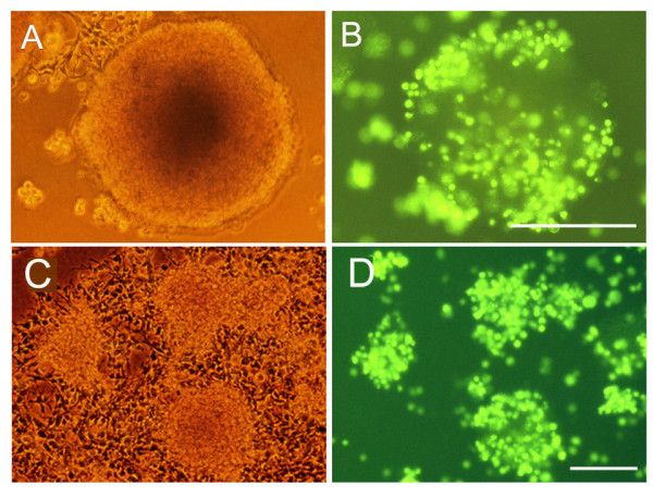
Brain EC attachment to neurospheres. Mouse brain EC were prepared, labelled with the fluorescent cell tracker marker (green), plated onto mouse neurospheres, and then cultured under serum-free conditions, as described in Methods. After 18 hours (A and B) and 5 days (C and D), EC adhesion to neurospheres was examined by fluorescent microscopy. Representative cultures are shown by phase (A and C) and fluorescent (B and D) images. Scale bars = 100 μm. Note that after 18 hours co-culture many fluorescent-labelled EC had attached to neurospheres and that after 5 days, many EC were still present, and were concentrated on or within neurospheres.
