
Fig. 3.
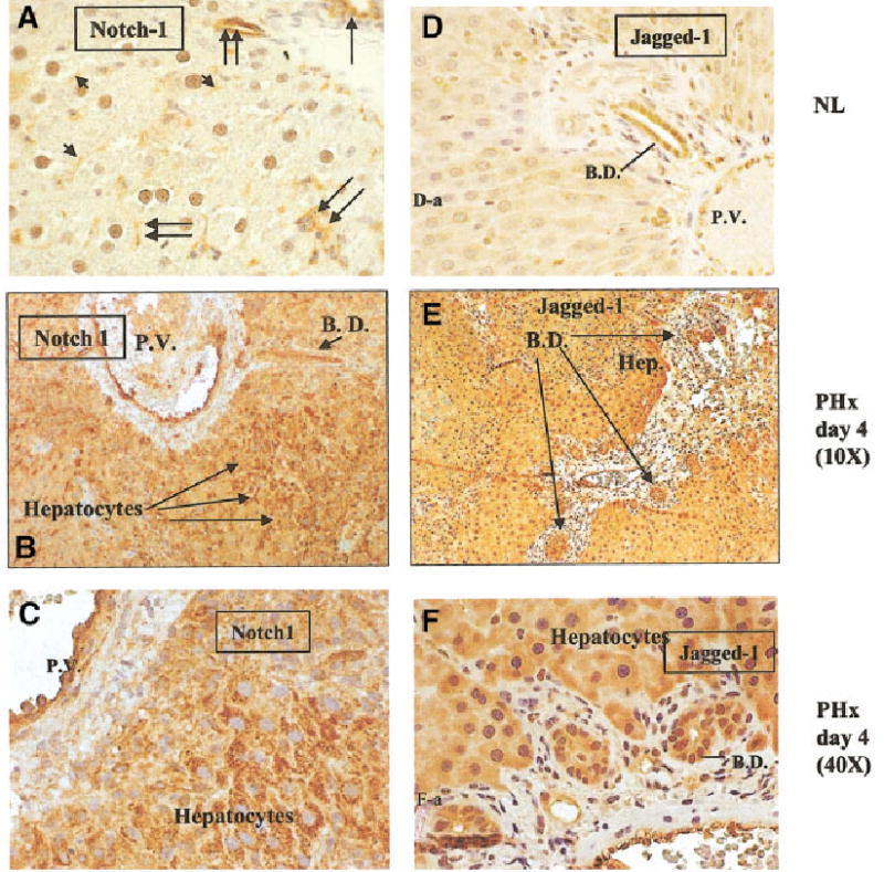
Time course of Notch and Jagged expression in regenerating liver determined by immunohistochemical staining. (A) Normal liver, magnification at 40×. Notch staining is shown on bile ductules (single long arrow), sinusoidal endothelial and small vessel endothelial cells (double arrows), and hepatocyte plasma membranes (short arrows). (B and C) Regenerating liver, 4 days after partial hepatectomy. Figure 3B was taken at a magnification of 10× and Fig. 3C at a magnification of 40×. Both pictures demonstrate staining of Notch in endothelial cells and periportal hepatocytes. (D) Normal liver (magnification: 20×). Jagged staining predominantly in bile ductules with weaker staining seen in hepatocytes. (E and F) Regenerating liver. Magnifications at 10× and 40×, respectively. Strong immunoreactivity for Jagged is seen in bile ductules and periportal hepatocytes. B.D. = bile ductile; P.V. = portal vein.