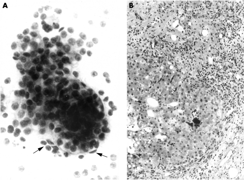
Figure 1.
Hepatocellular carcinoma. (A) Fine needle aspiration sample includes thickened trabeculae of hepatocytes with peripheral endothelial cells (arrows) and scattered atypical bare nuclei (above). Diff-Quik method, original magnification, ×300. (B) The corresponding core biopsy shows cirrhotic liver but no evidence of malignancy. Haematoxylin and eosin, original magnification, ×120.

