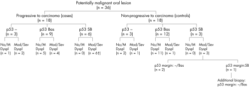
Figure 1.
Flow diagram depicting the most relevant characteristics and natural history of the potentially malignant oral lesions analysed. The p53 immunoexpression patterns of the lesions are shown in relation to their histopathological characteristics and behaviour upon follow up. Lesions that did not progress to carcinoma (controls) were selected by minimisation to match the group of lesions that progressed to carcinoma (cases) in variables such as patients' sex, age, and smoking habits, location of the lesion, and time of follow up. p53 immunostaining: −, negative; Bas, basal; SB, suprabasal. No/M Dyspl, no or mild dysplasia; Mod/Sev Dyspl, moderate or severe dysplasia; †suprabasal p53 extended into the margin of the section in five cases; p53 margin, p53 immunostaining at the margin of the section.

