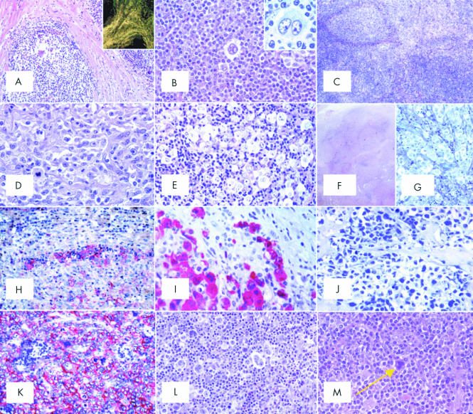
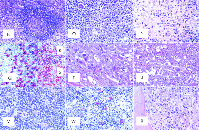
Figure 2.
(A) Nodular sclerosing common Hodgkin's lymphoma (NS-CHL): the normal lymph node structure is largely effaced because of a nodular growth; the nodule is surrounded by thick collagen bands originating from the capsule (haematoxylin and eosin; original magnification, ×40). Inset: birefringence of collagen bands (polarised light microscopy; original magnification, ×20). (B) NS-CHL: scattered lacunar cells can easily be seen within one nodule (haematoxylin and eosin; original magnification, ×300). Inset: cytological details of a lacunar cell (Giemsa; original magnification, ×800). (C) NS-CHL: so called cellular phase; note the nodularity of the growth (haematoxylin and eosin; original magnification, ×80). (D) NS-CHL: so called syncytial variant; note the cohesive growth pattern of neoplastic cells (haematoxylin and eosin; original magnification, ×400). (E) NS-CHL: an example of grade II tumour; note the content of neoplastic cells within the nodule, which covers more than 25% of the examined area (haematoxylin and eosin; original magnification, ×300). (F) An example of anaplastic large cell lymphoma of the Hodgkin-like type (ALCL-HL): the tumour consists of nodules partly surrounded by collagen bands (haematoxylin and eosin, original magnification, ×20).(G) ALCL-HL: at higher magnification it can be seen that the collagen bands are almost exclusively formed by neoplastic cells (Giemsa; original magnification, ×300). (H) ALCL-HL: typical intrasinusoidal diffusion of neoplastic cells as shown by CD30 staining (APAAP technique, Gill's haematoxylin counterstain; original magnification, ×200). (I) ALCL-HL: ALK protein expression by neoplastic cells (APAAP technique, Gill's haematoxylin counterstain; original magnification, ×400). (J) Primary mediastinal large B cell lymphoma (PMLBCL): neoplastic cells sometimes have multiple nuclei, show a wide rim of clear, fragile cytoplasm, and elicit a stromal reaction with compartmentalisation (Giemsa; original magnification, ×300). (K) PMLBCL: neoplastic cells express the CD30 molecule (APAAP technique, Gill's haematoxylin counterstain; original magnification, ×300). (L) Mixed cellularity common Hodgkin's lymphoma (MC-CHL): Hodgkin and Reed-Sternberg (H&RS) cells are easily identified; they are found with a cellular milieu consisting of small lymphocytes, some plasma cells, histiocytes, and granulocytes (haematoxylin and eosin; original magnification, ×350). (M) MC-CHL: an example of a mummified cell (arrow) (haematoxylin and eosin; original magnification, ×350). (N) MC-CHL: the tumour has a patent interfollicular location; a spared follicle with Castleman-like features can be seen (haematoxylin and eosin; original magnification, ×150). (O) MC-CHL: the tumour contains reactive epithelioid cells (haematoxylin and eosin; original magnification, ×300). (P) Peripheral T cell lymphoma not otherwise specified with a high content of epithelioid cells (so called Lennert's lymphoma): lymphoid elements show variation in size and shape (haematoxylin and eosin; original magnification, ×400). (Q) T cell rich, histiocyte rich large B cell lymphoma (TCRBCL): neoplastic cells strongly express the CD79a molecule (APAAP technique, Gill's haematoxylin counterstain; original magnification, ×300). (R) TCRBCL: reactive T cells and histiocytes are stained by the anti-CD3 antibody (APAAP technique, Gill's haematoxylin counterstain; original magnification, ×150). (S) TCRBCL: reactive T cells and histiocytes are stained by the anti-CD68 antibody (APAAP technique, Gill's haematoxylin counterstain; original magnification, ×150). (T) Lymphocyte depleted common Hodgkin's lymphoma (LD-CHL), fibrotic variant: rare neoplastic cells are surrounded by thick collagen bands with a haphazard organisation, some histiocytes and scanty lymphocytes (haematoxylin and eosin; original magnification, ×400). (U) LD-CHL, sarcomatous variant: H&RS cells are quite numerous; there is a certain degree of fibrotic reaction; small lymphocytes are exceedingly rare (haematoxylin and eosin; original magnification, ×350). (V) Lymphocyte rich common Hodgkin's lymphoma (LR-CHL): mononuclear and diagnostic neoplastic elements are found within a cellular milieu mostly consisting of small lymphocytes (haematoxylin and eosin; original magnification, ×350). (W) LR-CHL: neoplastic cells express CD15 (APAAP technique, Gill's haematoxylin counterstain; original magnification, ×300). (X) CHL: typical example of bone marrow involvement; note the fibrotic reaction, and the presence of H&RS cells (haematoxylin and eosin; original magnification, ×300).


