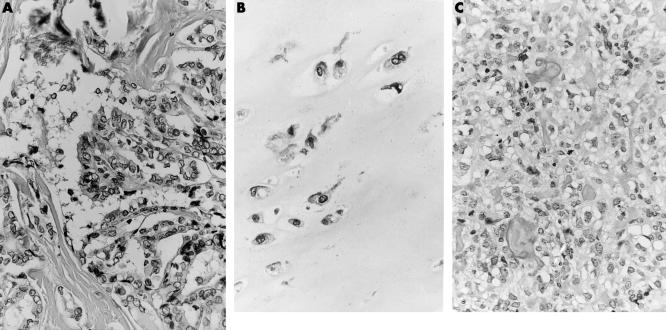
Figure 1.
(A) Light micrograph of the papillary thyroid carcinoma that was removed four years previously. (B) Light micrograph of the cartilaginous cap, displaying widely spaced, pronounced atypical, binucleated chondrocytes. (C) Light micrograph of a histologically distinct area present within the stalk of the tumour. This area is composed of an admixture of moderately pleomorphic tumour cells with the formation of a trabecular deposited osteoid. Note the presence of a mitotic figure.

