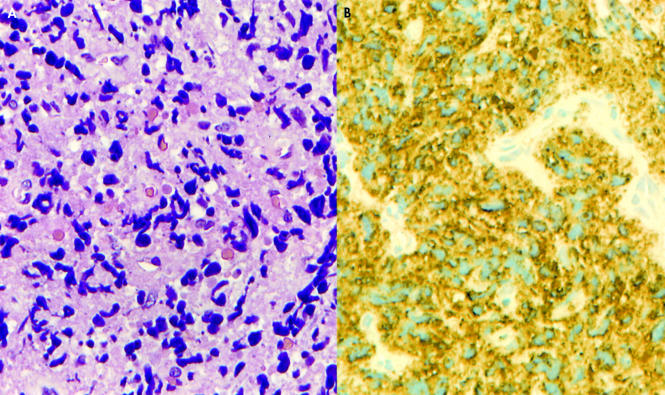
Figure 4.
(A) Initial biopsy specimen showing extensive crush artifact. Cell detail is absent and represented by streams of chromatin like material (haematoxylin and eosin stain; original magnification, ×400). (B) Anti-myeloperoxidase stain (original magnification, ×400) showing widespread positivity, confirming a diagnosis of extramedullary myeloid tumour.

