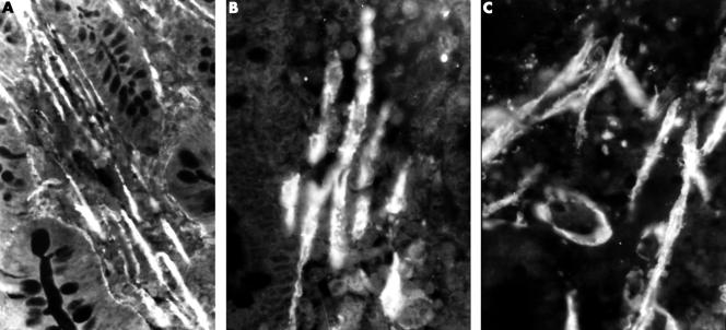
Figure 2.
Immunofluorescent staining for α-smooth muscle actin. (A) In healthy individuals, smooth muscle fibres appear as bundles of parallel elements between the crypts and in the axis of the villi; original magnification, ×100. (B) As the mucosa is damaged and villi become short and blunt the smooth muscle bands become less compact; original magnification, ×250. (C) When the mucosa is flat the smooth muscle fibres become disorganised and single fibres can be seen in any direction in the distal third of the mucosa; original magnification, ×200.

