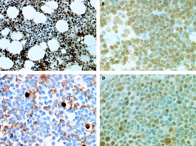
Figure 1.
The immunohistochemical patterns of expression of p33ING1b in normal tissue and acute lymphoblastic leukaemia (ALL). (A) Normal bone marrow showing nuclear expression of p33ING1b in almost all cells. (B) Normal thymus showing p33ING1b expression in precursor lymphoid cells. (C) ALL bone marrow trephine showing loss of p33ING1b from tumour nuclei with increase in cytoplasmic expression. (D) ALL bone marrow trephine showing p53 overexpression.

