Figure 1.
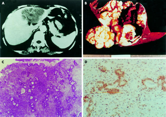
(A) Computed tomography scan showing a large cystic mass in the left lobe of the liver. (B) Left liver segment opened to show a large cystic mass. After rinsing away of the chocolate coloured content, the inner surface of the cyst appears yellow/white, uneven, and nodular. (C) Photomicrography of the cyst wall shows typical endometrial tissue with both glandular and stromal components (haematoxylin and eosin stained; original magnification, ×13.2). (D) Positive nuclear immunostaining with oestrogen receptor specific antibody in both glandular and stromal cells (DAB chromogen and haematoxylin counterstain; original magnification, ×66).

