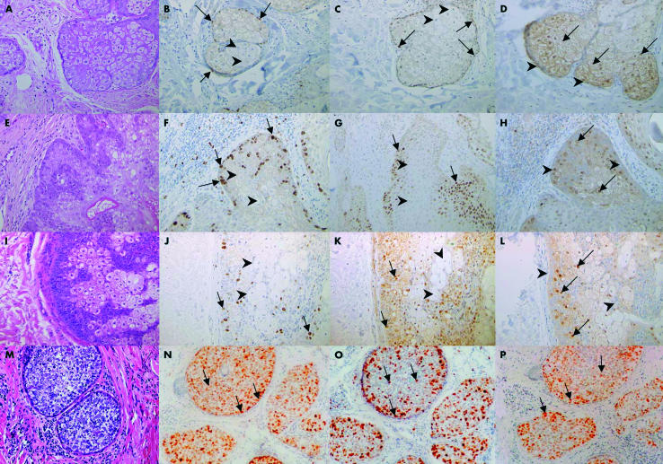
Figure 1.
(A–D) Normal sebaceous gland, (E–H) sebaceous adenoma, (I–L) sebaceoma, and (M–P) sebaceous carcinoma were stained with haematoxylin and eosin (A, E, I, M) or immunostained with antibodies to Ki67 (B, F, J, N), p53 (C, G, K, O), or p21WAF1 (D, H, L, P), as indicated. Normal sebaceous gland (A–D): basaloid cells show nuclear staining for Ki67 and p53 (arrows) but are negative for p21WAF1 (arrowheads). Differentiated sebocytes show nuclear staining for p21WAF1 (arrows) but are negative for Ki67 and p53 (arrowheads). Sebaceous adenoma (E–H) and sebaceoma (I–L): basaloid cells show increased nuclear staining for Ki67 and p53 (arrows) but are negative for p21WAF1 (arrowheads). Differentiated sebocytes show p21WAF1 nuclear positivity (arrows) but are negative for Ki67 and p53 (arrowheads). Sebaceous carcinoma (M–P) shows very disordered nuclear staining for Ki67, p53, and p21WAF1 (arrows), with a lack of compartmentalisation. The staining intensity is increased compared with normal glands and adenomas.

