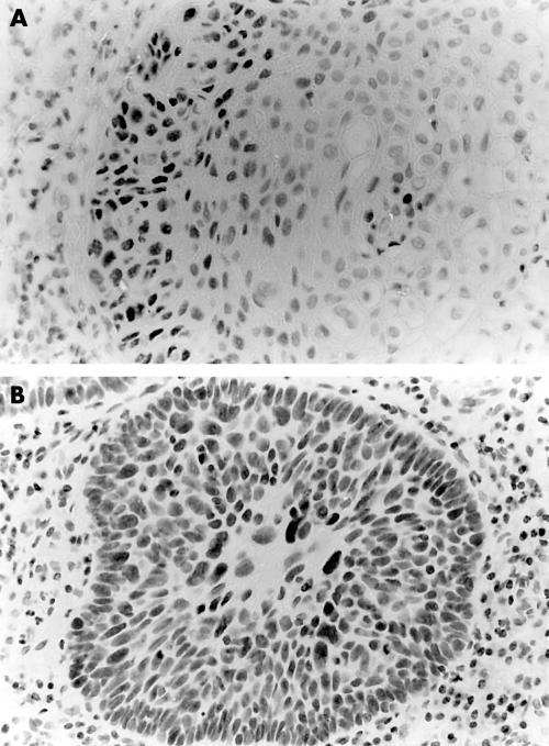
Figure 1.
(A) Oral squamous cell carcinomas (OSCC) showing p53 immunostaining in between 25% and 50% of the neoplastic cells (tables 1 and 2; T38). Note that mainly the peripheral layers of the tumour stains for p53 (p53 immunohistochemistry, counterstained with haematoxylin); (B) OSCC showing p53 immunostaining in almost all neoplastic cells (table 1; T53) (p53 immunohistochemistry, counterstained with haematoxylin).

