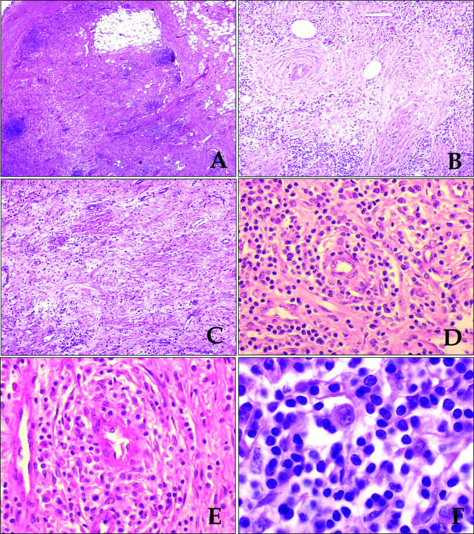
Figure 1.
(A) The lymph node had a hypocellular appearance with residual small lymphoid follicles. The capsule is shown on the left side. (B,C) Lymphoid cells were loosely dispersed in an oedematous stroma, rich in venules. (D,E) Higher magnifications showed small to medium sized lymphoid cells dispersed in a oedematous stroma, with accentuation of the cellularity around the venules. (F) Rare large cells with the features of hallmark cells (eccentrically located reniform or embryo-like nucleus and abundant amphophilic cytoplasm) were present.

