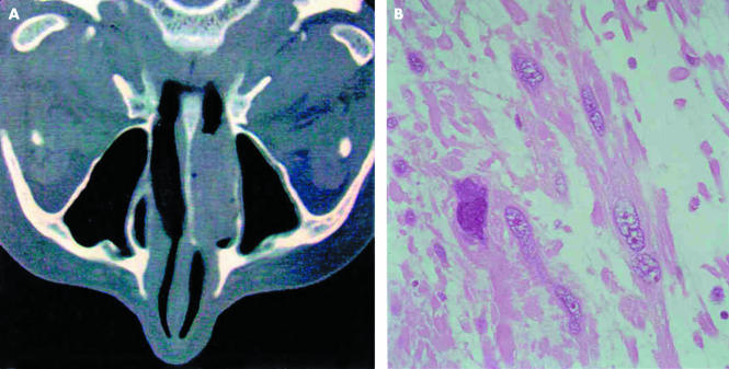
Figure 1.
(A) Axial computed tomography scan of the nasal sinuses shows a polypoid lesion occupying the whole inferior nasal fossa. (B) At histological examination, the tumour consisted of a proliferation of spindle shaped, pleomorphic cells with eosinophilic cytoplasm in a prominent myxoid background. Haematoxylin and eosin stained; original magnification, ×250.

