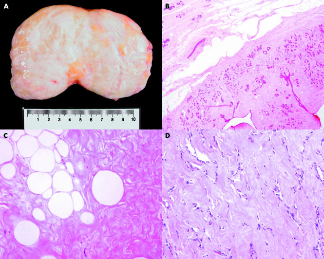
Figure 2.
(A) Specimen picture of a breast hamartoma. (B) Photomicrograph showing the smooth border of a hamartoma (haematoxylin and eosin stained; original magnification, ×20). (C) Photomicrograph showing single and small clusters of adipocytes within a densely fibrotic stroma (haematoxylin and eosin stained; original magnification, ×200). (D) Photomicrograph showing pseudo-angiomatous changes of the stroma (haematoxylin and eosin stained; original magnification, ×100).

