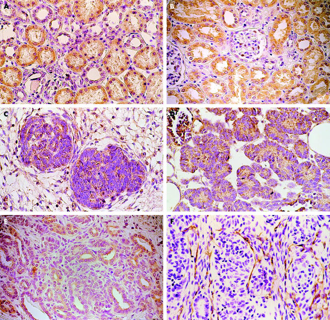
Figure 1.
Immunohistochemical staining for vascular endothelial growth factor (VEGF) and its receptor (Flt-1) in (A, B) normal renal tissue and (C–E) nephroblastoma tissue. (A) VEGF and (B) Flt-1 were detected in the tubular structures of normal renal tissue. VEGF and Flt-1 were mainly identified in the cytoplasm of the blastemal component (C and E, respectively) and in the epithelium of nephroblastoma tissue (D and E, respectively). (F) Nephroblastoma tissue stained with CD31 detected microvessels, which are seen in the stromal tissue. Original magnification, ×400.

