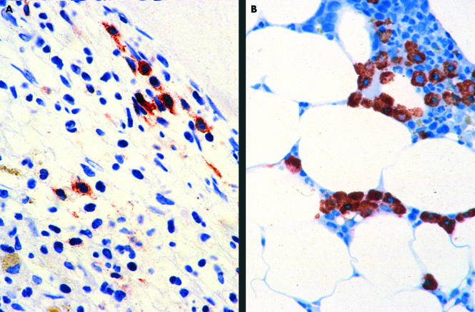
Figure 1.
Bone marrow in systemic mastocytosis. (A) An extremely hypercellular bone marrow in a case of aggressive systemic mastocytosis with an increase in spindle shaped mast cells. Very few of the mast cells are immunoreactive for chymase. (B) A hypocellular bone marrow with a focal increase in mast cells in a case of indolent systemic mastocytosis. Here, all the mast cells strongly express chymase. Avidin–biotin–peroxidase complex method, antichymase (B7) antibody.

