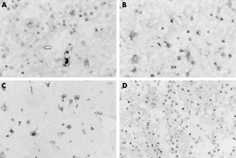
Figure 2.
Representative examples of vascular endothelial growth factor (VEGF) and pigment epithelium derived factor (PEDF) immunohistochemical staining. The positive signal of VEGF was most intense in endothelial cells, tumour cells, and vessels around areas of necrosis. The intensity of PEDF immunostaining decreased greatly from low grade glioma to high grade glioma. (A) VEGF expression in astrocytoma; (B) VEGF expression in glioblastoma; (C) PEDF expression in astrocytoma; (D) PEDF expression in glioblastoma (original magnifications, ×400).

