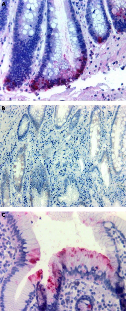
Figure 3.
Immunohistochemical detection of defensin peptides in selected gastric tissue sections. (A) Epithelial cells of the basal crypts stained positive for human defensin 5 (HD-5) with the alkaline phosphatase anti-alkaline phosphatase (APAAP) method (dilution of the polyclonal antiserum, 1/4000). (B) Human β defensin 1 (HBD-1) was detected by the peroxidase method (dilution of the polyconal antiserum, 1/1000) disclosing a broad epithelial distribution. (C) The APAAP method was performed for HBD-2 detection (dilution of the polyclonal antiserum, 1/2000). Positive cells were confined to the epithelial cells of the apical foveolae.

